An atlas of the spinal cord seeks to combat disease
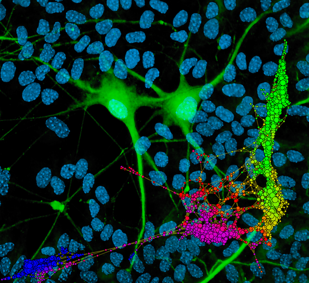
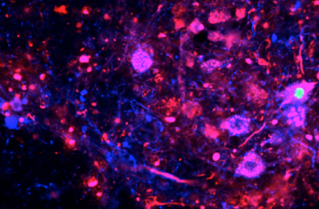
Central to our ability to walk, the spine also is the site of neurodegenerative diseases and traumatic injuries. To help develop effective treatments, scientists at Columbia’s Zuckerman Institute are mapping the spinal cord, cell by cell.
“We don’t understand all the cell types that exist within the brain and the spinal cord, and we also don’t understand their function,” says Abbas Rizvi, associate research scientist and leader of Columbia’s cell atlas project.
Now, the team is isolating individual cells from each segment of the spinal cord and performing genetic analysis on them with sophisticated computational tools. New imaging techniques are making it possible to peer inside human tissue to provide the most accurate look at cells in their environment. Together, these will create a highly detailed guide available to all. The project is supported by the Chan Zuckerberg Initiative.
“One of the things that I love the most about this project is that it really enables the construction of community, locally within our lab but also within our institute here at Columbia, and then even broader than that, interfacing with an international community,” says Rizvi. Theoreticians, mathematicians, and computational biologists across the University as well as around the world are working together to build this open resource.
In its pinpoint accuracy and accessibility, the guide holds tremendous potential for understanding and treating diseases like ALS. For example, in ALS, certain motor neurons are selectively vulnerable to cell death. With a deeper knowledge of these motor neurons’ function and location, scientists may be able to develop highly targeted therapies. Learn more.
Make Your Commitment Today
Ideas and Impact