Capturing images to improve CRISPR-based gene editing
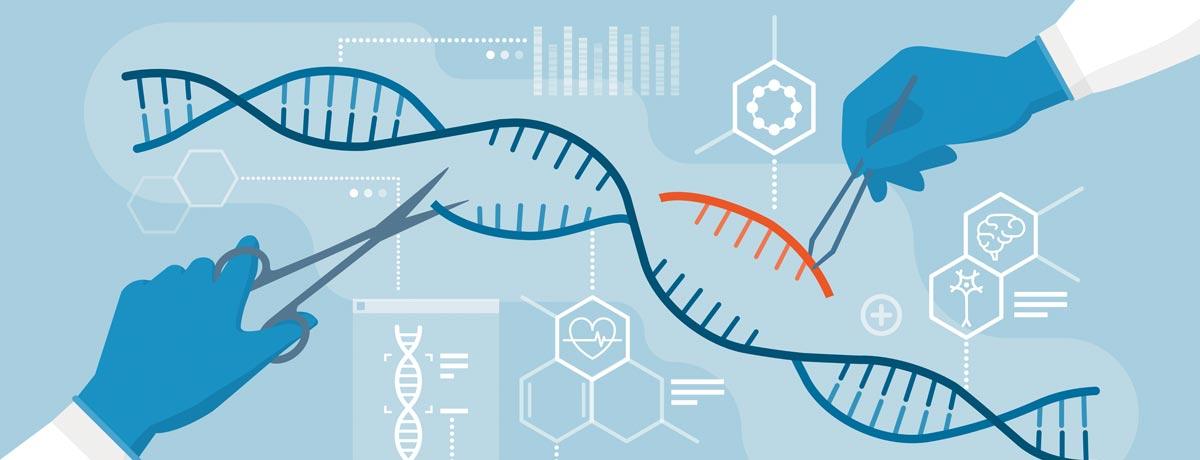
Using a Nobel prize winning technique called cryo-electron microscopy, Columbia researchers froze a new gene editing tool in action—and, for the first time, took pictures of it. These high-resolution images show how INTEGRATE, a new, CRISPR-based tool, actually works. And the pictures may help researchers build even more accurate gene editing tools, eventually improving biomedical treatments.
“These new images, a wonderful collaboration with Israel Fernández’s lab, explain the biology with incredible molecular detail and will help us improve the system by guiding protein engineering efforts,” said Sam Sternberg, assistant professor of biochemistry and molecular biophysics at Columbia’s Vagelos College of Physicians and Surgeons, who led the research with Fernandez, assistant professor of biochemistry and molecular biophysics.
The new gene editing tool, INTEGRATE, promises greater precision than existing, widely used CRISPR technologies. INTEGRATE, developed by the Sternberg lab, can insert large DNA sequences, for instance, without relying on the cell’s machinery to repair strands. This means INTEGRATE may be more efficient at gene editing than the original, well-known CRISPR-Cas system.
Thanks to clearer images of INTEGRATE at work, researchers plan to explore the new gene editing tool’s possibilities in treating diseases. Learn more.
Make Your Commitment Today
Ideas and Impact