Revolutionary microscope captures biology at the speed of life
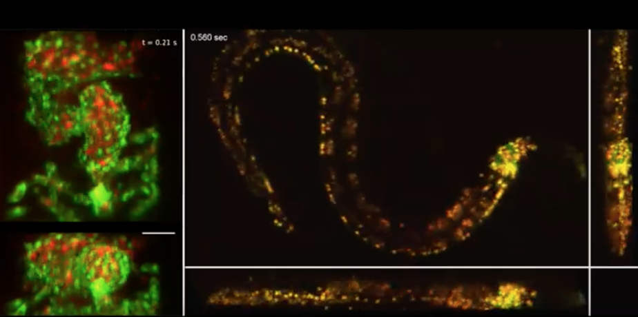
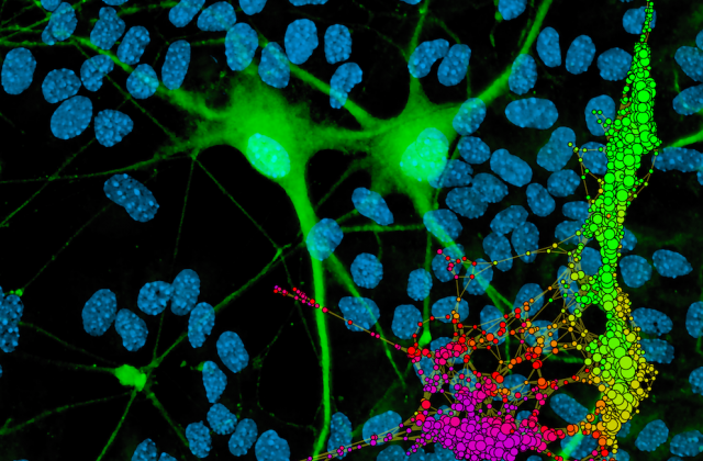
Columbia scientists have created a new high-speed microscope capable of observing the dynamic processes of living things, from the beating heart of a fish embryo to the wriggles of a worm. Called SCAPE 2.0, this new technology uses angled sheets of light to quickly scan entire planes, forming a 3D image.
SCAPE 2.0 improves on a technique conventional microscopes use to image living samples: scanning a small spot of laser light around the sample. This traditional approach is not only slower but also harder on living samples than SCAPE 2.0, since it uses much more light. Additionally, SCAPE 2.0 is smaller—it has a single stationary, objective lens rather than the multiple lenses other microscopes require. In fact, SCAPE 2.0 is so compact that Elizabeth Hillman, leader of the effort and a professor of biomedical engineering, often carries the system in the trunk of her car.
She has been giving hands-on demonstrations to help scientists around the world use SCAPE in their research. “SCAPE 2.0 opens up a new landscape of things we can see. I hope our new results will inspire scientists to think of what new questions can be asėed, and what new avenues of scientific discovery we can explore next.”
From imaging the way gas bubbles form in volcanic eruptions to tracking how genetic mutations affect heart development, SCAPE holds tremendous promise to transform a variety of fields. Learn more.
Make Your Commitment Today
Ideas and Impact